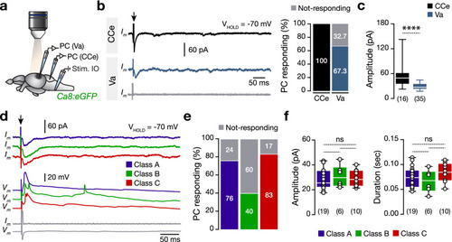
Direct electrical stimulation of inferior olive generates large-amplitude events in a proportion of valvular Purkinje cells. (a) Ex-vivo setup of an isolated intact brain from the Tg(Ca8:eGFP) line allows simultaneous whole-cell patch-clamp recordings of Purkinje cells and electrical stimulation of the inferior olive nucleus. (b) Voltage-clamp recordings of Purkinje cells showing the differential responses of the Purkinje cells located in corpus and valvula cerebelli. (c) Corpus cerebelli Purkinje cells responds to the inferior olive electrical stimulation, generating larger amplitude responses than the ones detected in the valvula cerebelli Purkinje cells. (d) Sample voltage- and current-clamp recordings show the responses of the adult valvular Purkinje cells to inferior olive stimulation pulse (Black arrow). Gray traces from a Purkinje cell that do not respond to inferior olive stimulation. (e) Quantification of the proportion of the valvular Purkinje cells that respond to the inferior olive stimulation. (f) Analysis of the recorded amplitude and duration between the Purkinje cells that respond to the inferior olive stimulation. IO inferior olive, PC Purkinje cell. Data are presented as box plots showing the median with 25/75 percentile (box and line) and minimum–maximum (whiskers). ns not significant. For detailed statistics, see Supplementary Table S1.
|
