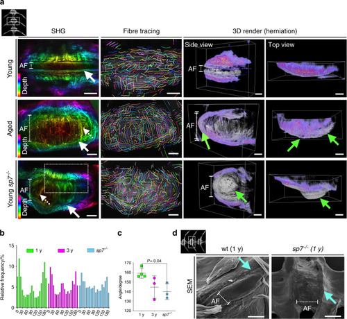
Altered collagen fiber organization and disc herniation in zebrafish IVDD. a Max projection of image stacks from second harmonic generation (SHG) imaging color coded by depth. Individual collagen fiber was traced using CTFire. 3D volumetric rendering from SHG images were used to visualize disc herniation in 3D using Amira (gray = back-scatter SHG; purple = forward-scatter SHG). Orientation of the vertebral column is shown at the top left of the panel. The annuls fibrosus (AF) is indicated with a dashed line. Note the variation in AF length among the groups. Smooth perpendicular fiber organization at the endplate was observed only in young samples (white arrow, SHG). Thicker fibers were observed in the AF of aged and sp7−/− zebrafish (dashed arrow). Bulging discs were found in aged zebrafish and severely bulging discs were found in sp7−/− zebrafish (green arrows, 3D render). Scale bars = 50 μm. b Relative frequency distribution (%) of angles of collagen fibers from each group. The graph was generated in Prism 8. c Angle of the most common collagen fibers (75% of total fibers) show abnormal orientation in sp7−/− fish (n = 3 fish per group). One-way ANOVA, post hoc Tukey’s multiple comparisons test; data are the mean and SD. P values are indicated when significant (P < 0.05). d Scanning electron microscopy (SEM) of 1-year-old wt and sp7−/− zebrafish (1 y). Note a smooth transition from the vertebral bone to the AF in wt zebrafish and a cliff in sp7−/− zebrafish (arrow). The orientation of the vertebral column is shown on the top left. Scale bars = 50 μm
|
