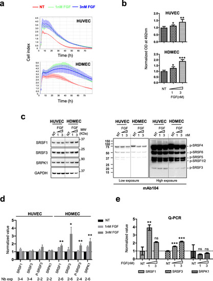
FGF-2 increases SRSF1, SRSF3 and SRPK1 protein levels in primary endothelial cells. a, b xCELLigence (a) or MTS (b) assay was used to assess HUVEC and HDMEC cellular adhesion, proliferation, and viability in response to FGF-2 at 1 or 3nM. Data are representative of at least 2 independent experiments performed in at least triplicate (mean ± SD, unpaired t test, **p<0.01; ***p<0.001). c Representative immunoblots for SRSF1, SRSF3, SRPK1 (left panel), and phospho-SR (p-SR) (mAb104, right panel, two exposure times) protein levels in HUVEC and HDMEC, respectively, treated with 1 or 3nM FGF-2 for 72 h. GAPDH was used as a loading control. NT: nontreated. d Semi-quantification using ImageJ software of SRSF1, SRSF3, P-SRSF3, or SRPK1 signal relative to GAPDH signal. Ratio obtained for NT group was arbitrarily assigned the value 1. Numbers below the graph indicate the number of biological replicates for each condition (mean ± SD, unpaired t test, *p<0.05, **p<0.01). e HDMEC were treated with 1 or 3nM FGF-2 as in (c). SRSF1, SRSF3, and SRPK1 mRNA levels were quantified by RT-qPCR in each condition. GAPDH was used as an internal control. Mean ± SEM are presented (n=4, unpaired t test, **p< 0.01, ***p<0.001, ns: not significant)
|
