
Figure 3
- ID
- ZDB-FIG-210902-251
- Publication
- Kurnia et al., 2021 - TCMacro: A Simple and Robust ImageJ-Based Method for Automated Measurement of Tail Coiling Activity in Zebrafish
- Other Figures
- All Figure Page
- Back to All Figure Page
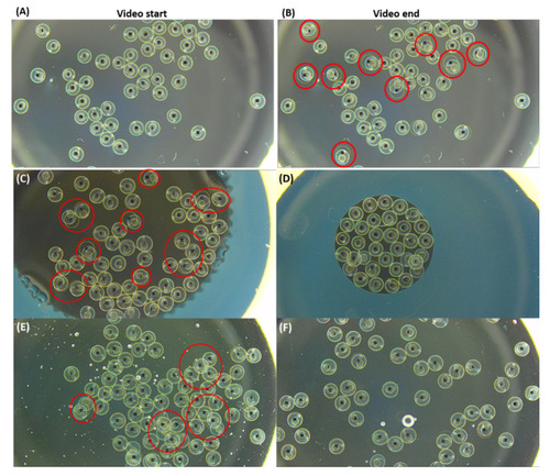
Comparison of different methods to reduce embryo position shifting during video recording for tail coiling measurement in zebrafish. (A) ROI selection in dissecting microscopy with darkfield setting. (B) Some embryos’ position was shifted due to embryo tail coiling (labeled by red circles). Two different sizes of agarose holders were tested to reduce the inter-embryo space with either (C) agarose outer wall (ø = 8.5 mm) or (D) agarose outer wall (ø = 4.4 mm). Two different concentrations of methylcellulose at either (E) 1% or (F) 0.5% were used to increase the mounting medium viscosity to prevent embryo position shifting. Egg movement caused by tail coiling occurrence was detected and marked with red circles. Images were captured at a low magnification at 6×. |