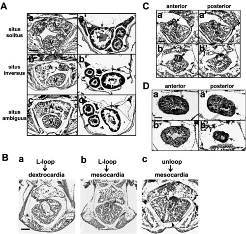
Histopathological assessment of heterotaxy in juvenile zebrafish. (A) Haematoxylin and eosin–stained transverse paraffin sections of 5-week-old zebrafish showing situs solitus, situs inversus, and situs ambiguus of the heart (a–c) and other visceral organs (a’–c’) in each fish. Atrium tip, red arrowhead; intestinal bulb, red arrow; two anterior and posterior intestines, black arrows. Malrotated guts (arrows) are shown in the fish. Scale bars, 100 µm. Laterality of visceral organs in the selected fish are summarized in Table 1. (B) L-looped heart in 48 hpf embryo developed to dextrocardia (a) or mesocardia (b) at 5 weeks of age, while unlooped heart developed to mesocardia (c). Additionally, some experimental fish had transposition of the bulbo-ventricular valve (b, red arrowhead), non-apex ventricle (c, black arrowhead), and abnormal atrioventricular canal (c, black arrow). Scale bar, 100 µm. (C) Abnormal structure and laterality defect of the outflow tract (bulbus arteriosus–ventricle) valve (black arrow), blood flow defect in the outflow tract (red arrowhead), and ectopic membrane (red arrow) are shown in Fish #12. Scale bar, 50 µm. (D) Coarctation of the outflow tract (arrow) with defective smooth muscle layer (arrowhead) was observed in Fish #12. Scale bar, 50 µm.
|
