|
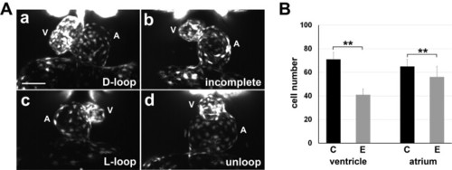
Cardiac looping defects and hypoplastic ventricle. (A) 3D SPIM imaging of the developing endocardium of Tg(fli1:EGFP) zebrafish embryos revealed cardiac defects in 50 hpf embryos from multiple in-crosses and cold shock: D-loop (a), incomplete loop (b), L-loop (c), and unloop (d) hearts. A, atrium; V, ventricle. Scale bar, 50 µm. (B) Total cell number in the endocardium of Tg(fli1:EGFP) zebrafish embryos at 50 hpf was counted using the 3D images (n = 6 of embryos with D-loop, n = 17 of embryos with laterality defects). Endocardial ventricles show more severe hypoplasia in the experimental embryos (E) compared with that in controls (C). Values are presented as mean ± SEM of total cell numbers. **P< .001 by two-sample Student’s t-test.
|