|
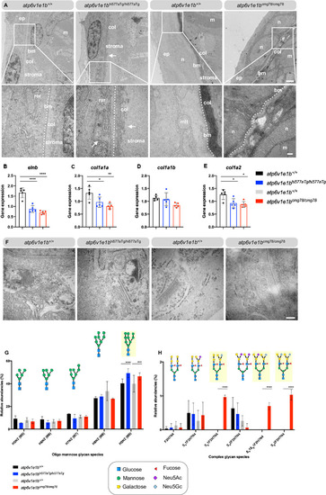
Profound epidermal and N-glycosylation alterations in <italic toggle='yes'>atp6v1e1b</italic>-deficient zebrafish.(A) Representative images of ultrathin sections taken from the dermis of atp6v1e1b-deficient zebrafish and WT controls at 4 dpf. Note the two-layered epidermis that is separated from the collagenous stroma of the dermis by a well-defined basement membrane (bm), indicated as dotted line, at this developmental timepoint. In atp6v1e1b-deficient larvae, we observed a larger and more disorganized collagenous stroma of the dermis (atp6v1e1bhi577aTg/hi577aTg) or folded bm (atp6v1e1bcmg78/cmg78). Reproducible results were obtained in three independent experiments. Scale bar = 1 μm (top panel), scale bar = 200 nm (bottom panel). Bm: basement membrane; col: collagenous fibrils; ep: epidermis; m: muscle cell surface; mit: mitochondria; n: nucleus; stroma: primary dermal stroma; rer: rough endoplasmic reticulum. (B-E) Quantification of elnb, col1a1a, col1a1b, col1a2 by RT-qPCR of atp6v1e1b-deficient larvae at 4 dpf compared to WT controls. Data are expressed as mean ± SD from 5 biological replicates. (F) Representative images demonstrating dilated Golgi apparatus in atp6v1e1bcmg78/cmg78. Reproducible results were obtained in three independent experiments. Scale bar = 200 nm (G) Quantification of relative abundancies of MALDI-TOF-MS signals for oligomannose N-glycan species detected in deyolked lysates in atp6v1e1b-deficient zebrafish compared to WT controls. (H) Quantification of relative abundancies of MALDI-TOF-MS signals for complex N-glycan species detected in deyolked lysates in atp6v1e1b-deficient zebrafish compared to WT controls. Data are expressed as mean ± SD from 3 biological replicates. 2-way ANOVA with Tukey test for multiple comparison was used for statistical analysis. Yellow shading indicates differences between WT controls and atp6v1e1b-deficient larvae. Symbols represent monosaccharide residues. Graphical representation is based on the accepted convention from the Symbol Nomenclature for Glycans (Consortium for Functional Glycomics). Yellow circle: Gal; green circle: Man; red triangle: Neu5Ac; light blue diamond: Neu5Gc; Neu5Ac: N-acetylneuraminic acid; Neu5Gc: N-glycolylneuraminic acid.
|