FIGURE

Fig. 1

ID
ZDB-FIG-210619-6
Publication
Zou et al., 2021 - In vivo imaging reveals mature Oligodendrocyte division in adult Zebrafish
Other Figures
All Figure Page
Back to All Figure Page
Fig. 1

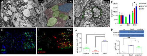
Oligodendrocytes survived after optic nerve injury. a At 1 dpi, axons degenerated in the distal stump, inducing myelin collapse. b At 7 dpi, microglia/macrophages (pink) ingested myelin debris (green). c Remyelination (green) started to ensheath regenerated axons at 7 dpi. d Statistical analysis of olig2+ cells in the proximal segment, epicenter, and distal segment at different times after injury. e-f At 5 dpi, olig2+ cells were not found in the epicenter. Most olig2+ cells in the distal stump (right side) expressed p-erk1/2 (yellow cells), while the proximal cells (left side) did not. g The percentage of p-erk1/2 in olig2+ cells in the distal segments was significantly higher than that in the proximal segments at 5 dpi. h Compared with the expression level of p-erk1/2 protein in the normal optic nerve, the proximal segments and the distal segments were at 5 dpi by western. Scale bar: 200 nm (a-c), 20 μm (f).

Expression Data

Expression Detail
Antibody Labeling
Phenotype Data

Phenotype Detail
Acknowledgments
This image is the copyrighted work of the attributed author or publisher, and ZFIN has permission only to display this image to its users. Additional permissions should be obtained from the applicable author or publisher of the image. Full text @ Cell Regen (Lond)