
Figure 5
- ID
- ZDB-FIG-210607-35
- Publication
- Li et al., 2021 - Abortive intussusceptive angiogenesis causes multi-cavernous vascular malformations
- Other Figures
-
- Figure 1
- Figure 1 - figure supplement 1
- Figure 1 - figure supplement 2
- Figure 2
- Figure 3
- Figure 3 - figure supplement 1
- Figure 4
- Figure 4 - figure supplement 1
- Figure 4 - figure supplement 2
- Figure 5
- Figure 5 - figure supplement 1
- Figure 6
- Figure 6 - figure supplement 1
- Figure 7
- Figure 7 - figure supplement 1
- Figure 7 - figure supplement 2
- All Figure Page
- Back to All Figure Page
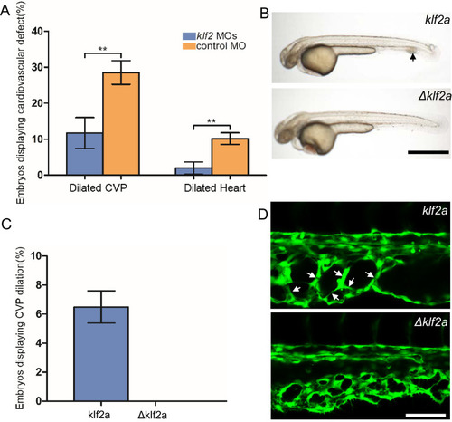
(A) Both the CVP dilation and heart dilation were rescued by injection of klf2 morpholinos in 2 days post fertilization (dpf) ccm2 CRISPR embryos. **p<0.01. Error bars indicate SD. (B) pCS2-KLF2a linearized DNA-injected 2.5 dpf embryos displayed CVP dilation, whereas injection of a DNA fragment containing a DNA binding domain deleted ΔKLF2a mutant showed normal development. Arrow indicates the CVP dilation and retained erythrocytes. Scale bar: 1 mm. (C) Quantification of the prevalence of CVP dilation following KLF2a or ΔKLF2a overexpression. The mean and SD are shown. (D) Representative images show the honeycombed lumen and dilated CVP in 1.5 dpf KLF2a-injected embryo and normal CVP of ΔKLF2a-injected embryo. Arrow indicates honeycombing. Scale bar: 100 µm. |
| Fish: | |
|---|---|
| Knockdown Reagents: | |
| Observed In: | |
| Stage: | Long-pec |