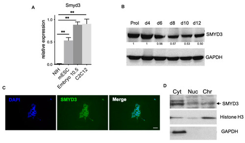
SMYD3 is expressed in mouse embryo and in mouse embryonic stem cells (mESCs). (A) mRNA was extracted from NIH3T3, C2C12 myoblasts and mESC in parallel with d10.5 embryo and Smyd3 transcript levels were measured by qRT-PCR. Data were normalized to GAPDH. Means from three independent experiments are shown. ** p < 0.01 value for the significance is shown in the plot. (B) Immunoblot analysis of SMYD3 protein levels at different time points of ESC differentiation. GAPDH served as a loading control. Normalized band intensity in immunoblots is reported below signals. Representative image of three independent experiments. (C) Immunofluorescence was performed on undifferentiated mESCs with an antibody raised against SMYD3. Nuclei were visualized by DAPI staining (blue). Scale bar: 50 μm. (D) ESCs were analyzed by biochemical fractionation, followed by immunoblot, to characterize SMYD3 distribution in different fractions: cytoplasmic or soluble fraction (Cyt), solubilized nuclear proteins fraction (Nuc), and chromatin-nuclear matrix-bound fraction (Chr). GAPDH served as Cyt control, while H3 served as Chr control. Representative image of two independent experiments.
|
