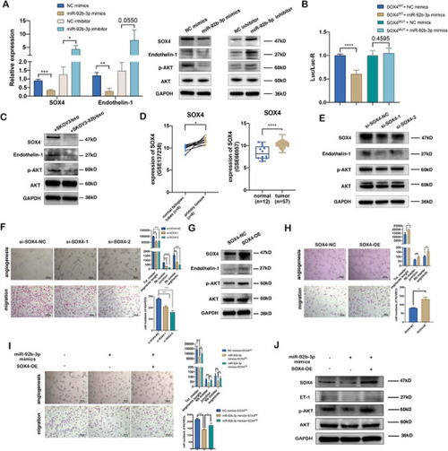
SOX4 is a target of exosomal miR‐92b‐3p and participates in exosomal miR‐92b‐3p modulating angiogenesis. (A) qRT‐PCR analysis (left) and Western blotting (right) of the relative expressions of SOX4, endothelin‐1, and p‐AKT in HUVECs transfected with miR‐92b‐3p mimics or miR‐92b‐3p inhibitor (mean ± SD, n = 3). (B) Dual luciferase reporter of verifying the combination between 3′UTR of SOX4 gene and miR‐92b‐3p. The pmirGLO reporters containing 3′UTR of human SOX4 gene with wild‐type (wt) or mutated (mut) miR‐92b‐3p binding sites were used to transfect HUVECs while treating with miR‐92b‐3p mimics or NC mimics (control). Luciferase activity was analyzed at 48 h post transfection (n = 6 extracts) and the ratio between Renilla luciferase and firefly luciferase activities (Rluc/Fluc) is shown. (C) Western blotting of the expression level of SOX4, endothelin‐1, and p‐AKT in HUVECs treated with SKOV3/exo and SKOV3‐92b/exo, respectively. (D) Relative expression of SOX4 in ovarian tumors compared to the epithelium of the normal fallopian tubes (GSE137238 data, left) and normal ovarian tissues (GSE66957 data, right). (E) Western blotting of the expressions of SOX4, endothelin‐1, and p‐AKT in HUVECs transfected with two siRNAs of SOX4 (si‐SOX4‐1, si‐SOX4‐2). (F) Representative images of tube formation and migration of HUVECs treated with si‐SOX4‐1 or si‐SOX4‐2 (scale bar = 100 μm). Total master segments length, number of master junctions, number of master nodes, and migration cell numbers were regarded as indicators of angiogenic ability in vitro and assessed by ImageJ (mean ± SD, n = 3). (G) Western blotting of the expressions of SOX4, endothelin‐1, and p‐AKT in HUVECs transfected with SOX4‐overexpression plasmids. (H) Representative images of tube formation and migration of HUVECs treated with SOX4‐overexpression plasmids (scale bar = 100 μm). Total master segments length, number of master junctions, number of master nodes, and migration cell numbers were regarded as indicators of angiogenesis ability in vitro and assessed by ImageJ (mean ± SD, n = 3). (I) Representative images of tube formation and migration of HUVECs treated with miR‐92b‐3p mimics or miR‐92b‐3p mimics plus SOX4‐plasmids. Total master segments length, number of master junctions, master nodes, and migration cells were regarded as indicators of angiogenic ability in vitro and assessed by ImageJ (mean ± SD, n = 3) (scale bar = 100 μm). (J) Western blotting of SOX4, endothelin‐1, and p‐AKT proteins in HUVECs cotransfected with miR‐92b‐3p mimics plus SOX4 plasmids. Data are shown by at least three independent experiments, and the Student's t‐test was used to compare differences. *p < .05, **p < .01, ***p < .001, ****p < .0001
|
