
Fig. 1
- ID
- ZDB-FIG-210526-52
- Publication
- Hu et al., 2021 - An Arrhythmic Mutation E7K Facilitates TRPM4 Channel Activation via Enhanced PIP2 Interaction
- Other Figures
- All Figure Page
- Back to All Figure Page
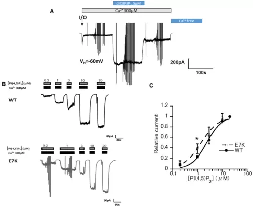
E7K mutant presented a higher PI(4,5)P28-PI(4,5)P2 (5 μM) restored TRPM4 channel activity over its initial magnitude. Vertical deflections reflect ramp- or step pulse-induced currents. (B) Representative data from two inside-out patch membranes (WT and E7K) demonstrating the dose-dependent reactivation of TRPM4 channels in response to various concentrations of diC8-PI(4,5)P2. (C) The degree of reactivation by diC8-PI(4,5)P2 (relative current) is defined as (I[x] − I[post-desens])/(I[20μM diC8-PIP2] − I[post-desens]), where I[x] and I respectively denote the amplitudes of TRPM4 currents reactivated by given (‘X’ μM) and maximal (20 μM) concentrations of diC8-PI(4,5)P2, and I[post-desens] is that of the basal TRPM4 current after desensitization to 300 μM Ca2+ [13]. Averaged concentration-response curves for the TRPM4 channel reactivation by diC8-PI(4,5)P2 are fitted by the Hill-type equation: 1/(1 + (EC50/[diC8PIP2])n. It gives EC50 values of 2.40 ± 0.23 μM and 1.44 ± 0.20 μM; Hill coefficient (n) values of 1.5 and 1.2 for WT and E7K, respectively. * p < 0.05 with ANOVA followed by Tukey’s post hoc tests (n = 5). |