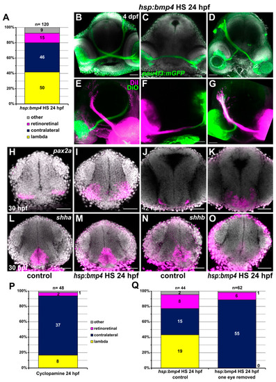
Late induction of bmp4 at 24 hpf results in different RGC projection defects. (A) Chart showing the distribution of phenotypes in tg(hsp70l:bmp4) embryos after heat shock at 24 hpf. Controls had exclusively contralateral projections (n = 78). (B–G) RGC projection phenotypes in tg(hsp70l:bmp4) embryos after heat shock at 24 hpf. (B–D) Immunohistochemistry against GFP in tg(pou4f3:mGFP), DAPI counterstaining, 4 dpf. (E–G) Injection of DiI/DiO, 3 dpf. (B,E) contralateral phenotype, (C,F) retinoretinal phenotype, (D,G) lambda phenotype. (H–O) In situ hybridization of in (I,K,M,O) tg(hsp70l:bmp4) embryos and (H,J,L,N) controls after heat shock at 24 hpf. (H,I) pax2a, 30 hpf; (J,K) pax2a, 42 hpf; (L,M) shha, 30 hpf; (N,O) shhb, 30 hpf, DAPI counterstaining. (P) Chart showing the distribution of phenotypes in embryos treated with 100 µM Cyclopamine at 24 hpf. Controls had exclusively contralateral projections (n = 30). (Q) Chart showing the distribution of phenotypes in tg(hsp70l:bmp4) embryos after heat shock at 24 hpf and subsequent removal of one eye, and in controls. All images are maximum intensity projections, transverse view, scale bars 50 µm.
|
