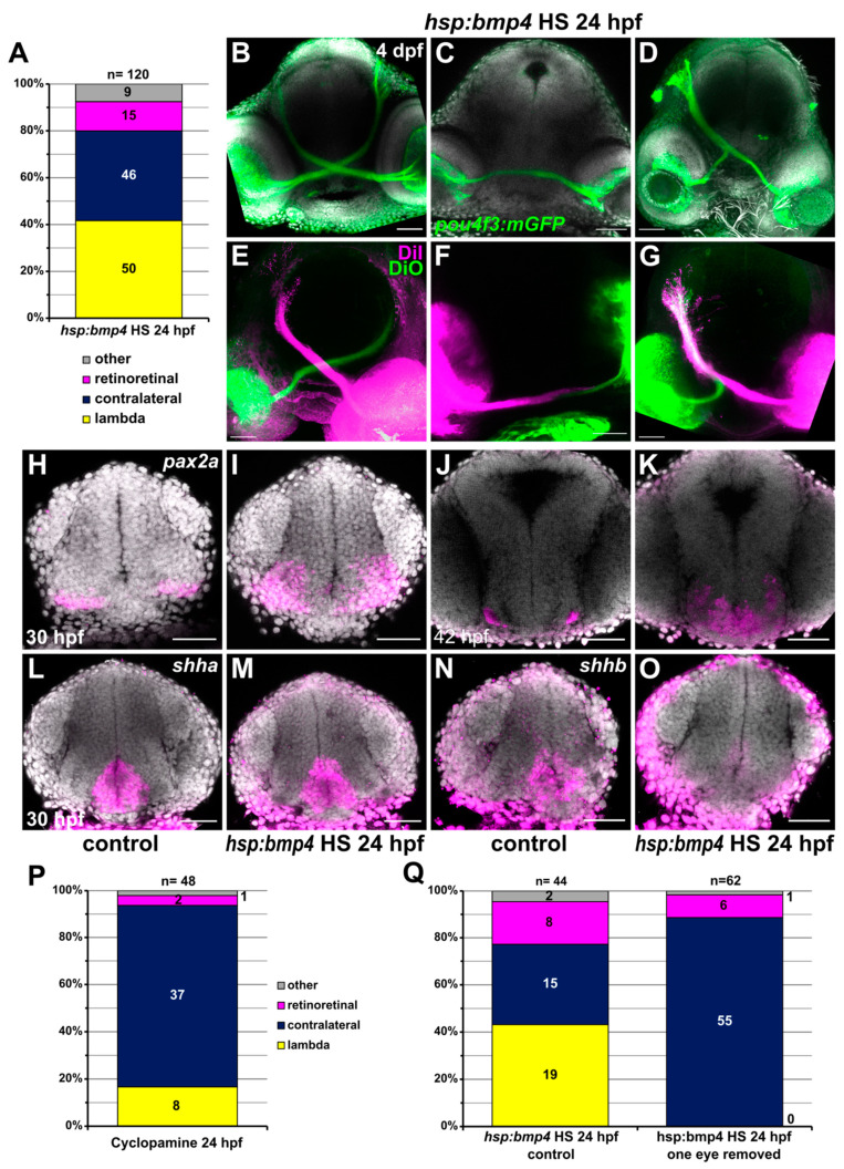
IMAGE

Figure 6

ID
ZDB-IMAGE-210519-79
Source
Figures for Knickmeyer et al., 2021
Image
Figure Caption

Figure 6

Late induction of bmp4 at 24 hpf results in different RGC projection defects. (A) Chart showing the distribution of phenotypes in tg(hsp70l:bmp4) embryos after heat shock at 24 hpf. Controls had exclusively contralateral projections (n = 78). (BG) RGC projection phenotypes in tg(hsp70l:bmp4) embryos after heat shock at 24 hpf. (BD) Immunohistochemistry against GFP in tg(pou4f3:mGFP), DAPI counterstaining, 4 dpf. (EG) Injection of DiI/DiO, 3 dpf. (B,E) contralateral phenotype, (C,F) retinoretinal phenotype, (D,G) lambda phenotype. (HO) In situ hybridization of in (I,K,M,O) tg(hsp70l:bmp4) embryos and (H,J,L,N) controls after heat shock at 24 hpf. (H,I) pax2a, 30 hpf; (J,K) pax2a, 42 hpf; (L,M) shha, 30 hpf; (N,O) shhb, 30 hpf, DAPI counterstaining. (P) Chart showing the distribution of phenotypes in embryos treated with 100 µM Cyclopamine at 24 hpf. Controls had exclusively contralateral projections (n = 30). (Q) Chart showing the distribution of phenotypes in tg(hsp70l:bmp4) embryos after heat shock at 24 hpf and subsequent removal of one eye, and in controls. All images are maximum intensity projections, transverse view, scale bars 50 µm.

Acknowledgments
This image is the copyrighted work of the attributed author or publisher, and ZFIN has permission only to display this image to its users. Additional permissions should be obtained from the applicable author or publisher of the image. Full text @ Int. J. Mol. Sci.