|
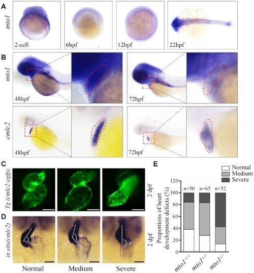
Heart development defects in zebrafish. (A) Whole-mount in situ hybridization (WISH) analysis of mto1 expression on wild type larval zebrafish at various ages (2-cell to 24 hpf). (B) WISH analysis of mto1 on wild type larval zebrafish at 48 hpf and 72 hpf. cmlc2 marker was used to indicate the place of heart. Insets show higher magnifications of heart. (C) Tg (cmlc2:egfp) embryos at 2 dpf shows the heart-restricted GFP expression in both chambers. Scale bars: 50 μm. (D) WISH against cmlc2. According to the looping state, measured by the angles between ventricle and atrium, hearts were divided into normal (α < 90°), medium (90° < α < 180°), and severe (α > 180°). Scale bars: 50 μm. (E) The proportions of each phenotypes in the embryos in mutant and WT zebrafish.
|