
Figure 1.
- ID
- ZDB-IMAGE-210512-28
- Genes
- Publication
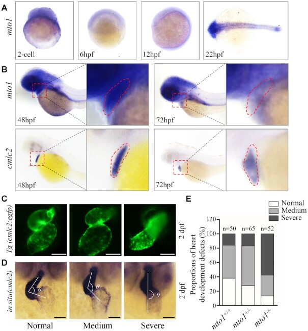
- Zhang et al., 2021 - Ablation of Mto1 in zebrafish exhibited hypertrophic cardiomyopathy manifested by mitochondrion RNA maturation deficiency
- All Figures
- Figures for Zhang et al., 2021
|
Figure 1.
Heart development defects in zebrafish. (