
Figure 3
- ID
- ZDB-FIG-210503-188
- Publication
- Pfefferli et al., 2021 - Persistent Ventricle Partitioning in the Adult Zebrafish Heart
- Other Figures
- All Figure Page
- Back to All Figure Page
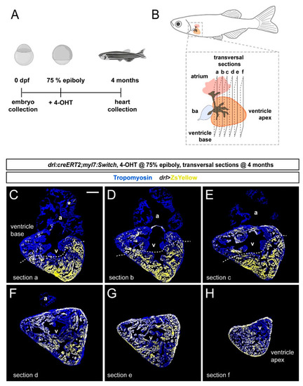
FHF contribution to the adult zebrafish ventricle remains localized. (A) Schematic of the experimental design and timeline. Embryos were 4-OHT-treated at 75% epiboly and raised to adulthood for 4 months. (B) Hearts were dissected from the adult animals and sequentially sectioned from the base of the heart to the apex as per schematic. (C–H) Series of transversal sections of a selected heart, immunostained against Tropomyosin (blue) and showing ZsYellow expression (yellow) from drl:creERT2-recombined myl7:Switch with atrium (a) on top and ventricle (v) at the bottom. Asterisk denotes rare atrial ZsYellow clones throughout sections. Note the markedly larger area of ZsYellow-labeled cardiomyocytes in the ventricle. Scale bar: 200 µm. |