
Figure 2
- ID
- ZDB-FIG-210503-187
- Publication
- Pfefferli et al., 2021 - Persistent Ventricle Partitioning in the Adult Zebrafish Heart
- Other Figures
- All Figure Page
- Back to All Figure Page
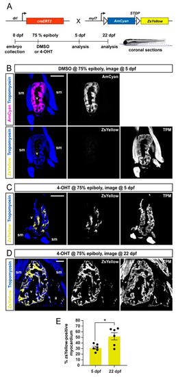
FHF lineage labeling persists during developmental stages. (A) Crossing scheme of used transgenes and timeline of experimental setup with 5 days post-fertilization (dpf) embryos and 22 dpf larvae longitudinally sectioned for heart analysis. (B–D) Confocal images of individual coronal sections, stained for pan-muscle Tropomyosin (blue, sm indicating skeletal muscles, v indicating ventricle, a indicating atrium). Embryos at 5 dpf (B), treated with DMSO (control, n = 4) at the 75% epiboly, show only myocardial AmCyan expression from myl7:Switch, while 4-OHT treatment (C) results in ZsYellow-expressing clones from drl:creERT2 activity, as also observed at 22 dpf (D). (E) Quantification of the ZsYellow–Tropomyosin double-positive cardiomyocytes of analyzed ventricles, two-tailed Student’s t-test, * p = 0.0149, 5 dpf n = 6; 22 dpf n = 6. Scale bar 50 µm. |