
Figure 7
- ID
- ZDB-FIG-210503-128
- Publication
- Lai et al., 2021 - The Great Capacity on Promoting Melanogenesis of Three Compatible Components in Vernonia anthelmintica (L.) Willd.
- Other Figures
- All Figure Page
- Back to All Figure Page
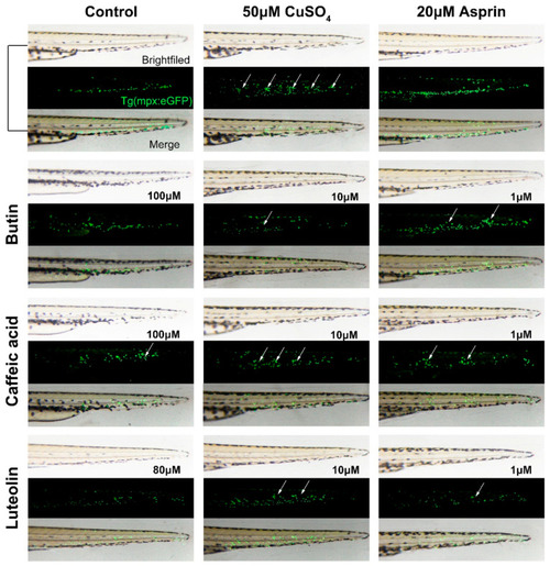
The anti-inflammatory effect of compounds in the chemically induced inflammation model. The time of image acquisition was when Tg(mpx:GFP) zebrafish larvae develop to 56 hpf. Currently, transgenic zebrafish larvae exhibit green fluorescent leukocytes. Untreated fish (control) show the normal distribution of labeled cells, mostly localized in the ventral trunk and tail. In copper-treated siblings (50 μM CuSO4), leukocytes become localized preferentially to a few clusters along the horizontal midline of the trunk and tail (see white arrows); no overt tissue damage to the larvae is observed in bright-field images., and relieving inflammation by applying aspirin (20 μM) as a positive standard. Before treated with CuSO4, zebrafish larvae were incubated with butin and caffeic acid [1, 10, 100] μM. |