|
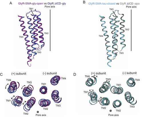
Conformational changes in the transmembrane domains of the zebrafish full-length GlyR andGlyR ΔICD. The full-length GlyR and the GlyR ΔICD are in the open (A and C) and closed (B and D) states, respectively. A and B, superposition of transmembrane domains from a single subunit from the full-length GlyR and the GlyR ΔICD from lateral view. C and D, superimposition of the (–)subunits illustrates the relative movements in the transmembrane domain of the (+)subunit. The view is from the intracellular side. GlyR full-length structures from Yu et al. (57), glycine bound open, PDB ID code 6PM6; taurine bound closed, PDB ID code 6PM3. GlyR ΔICD structures from Du et al. (2), glycine bound open, PDB ID code 3JAE; closed, PDB ID code 3JAD.
|