
Figure 6
- ID
- ZDB-IMAGE-210413-10
- Publication
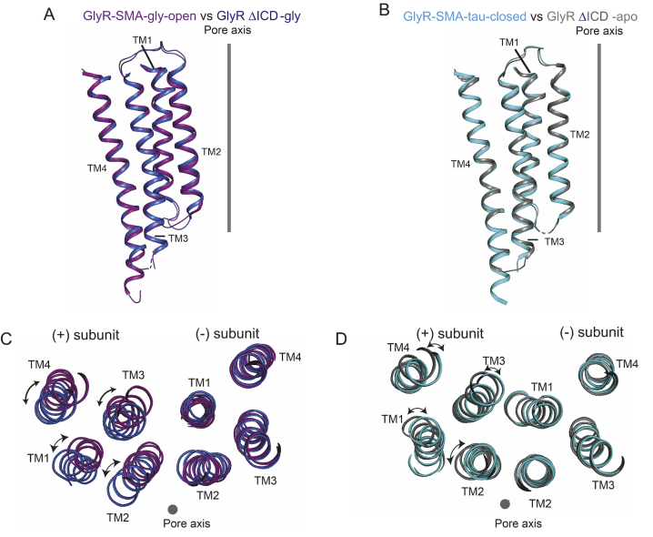
- Ivica et al., 2021 - The intracellular domain of homomeric glycine receptors modulates agonist efficacy
- All Figures
- Figures for Ivica et al., 2021
|
Figure 6