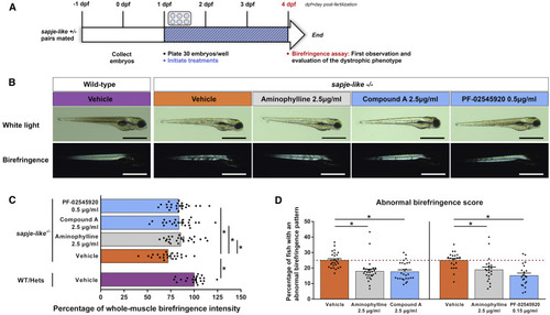
Short-Term Birefringence Screening Assay in Dystrophin-Deficient Zebrafish Identifies Phosphodiesterase 10A (PDE10A) Inhibitors That Reduce the Manifestation of the Dystrophic Muscle Phenotype (A) Design of short-term birefringence screening assay in dystrophin-deficient sapje-like zebrafish: pairs of sapje-like+/− zebrafish were mated and treatments were applied to the progeny in six-well plates (30 embryos per well) from 1 day post-fertilization (dpf). At 4 dpf, a birefringence assay was performed. (B) White light and birefringence images of representative 4-dpf larvae treated with 0.1% DMSO (vehicle, control), 2.5 μg/mL aminophylline (positive control), 2.5 μg/mL compound A, or 0.5 μg/mL PF-02545920 (two PDE10A inhibitors from Pfizer). Scale bars represent 1 mm. (C) Dot plots showing percentage of whole-muscle birefringence intensity of 4-dpf larvae. The whole-muscle birefringence intensity was normalized to the area of measure and larvae were genotyped. At least 22 (n = 22) zebrafish per experimental treatment were analyzed in three (N = 3) independent experiments. Statistical differences between groups are presented as follows: ∗p < 0.05 (t test, ±SEM). WT, wild-type larvae; Hets, heterozygous sapje-like+/− larvae. (D) Dot plots showing percentage of 4-dpf larvae that display abnormal muscle birefringence pattern (named as abnormal birefringence score) in each well. Mendelian genetics dictates 25% of the progeny to be dystrophin null and exhibit the muscle dystrophic phenotype seen by birefringence (red dashed line). Experimental treatments were performed in triplicate wells in at least seven (N = 7) independent experiments. Statistical differences between groups are presented as follows: ∗p < 0.05 (t test, ±SEM).
|
