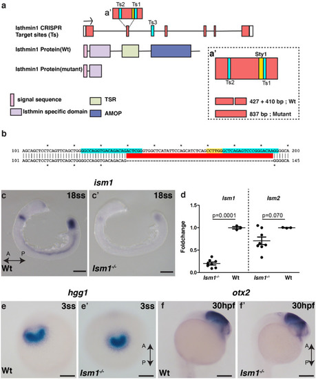
Generation and characterization of ism1 mutants a CRISPR gRNA target sites (Ts) were located in the exon (Ts1 and Ts2) and in the intron (Ts3). The sty1 restriction site is marked in yellow. Scheme representing protein domains in wild-type Ism1 and in the truncated version in CRISPR mutants are shown. Schematic representation of the genotyping strategy shown in a′, loss of the Sty1 restriction site in the mutants will result in an 837-bp band, while in the Wt there will be two bands (i.e., 437 + 410). b Sequence alignment between Wt and Ism1 mutant embryos shows 55 bp deletion, in cyan (Ts), and yellow (Sty1 restriction site). c and c′ Whole mount in situ hybridization in 18 somite stage (18ss; 18 hpf) embryos for Ism1 shows decreased expression in Ism1−/− embryos compared with the wild type (Wt) embryos. d qRT PCR for Ism1 and Ism2 in 24 hpf embryos of ism1−/− mutants show a significant reduction in only Ism1 mRNA with no change in Ism2 levels. e and e′ Whole mount in situ hybridization for hgg1, a marker for the anterior prechordal plate and the later hatching gland, shows similar expression and distribution patterns in the hatching gland between Wt and Ism1−/− embryos. Ventral images are shown. f and f′ Midbrain marker otx2 shows no obvious difference between Wt and Ism1−/− embryos. The anterior–posterior axis of the embryo is marked as A-P with an arrow. The total number of embryos n analyzed in panels c (n = 20), d (ism1−/−: n = 8, Wt: n = 3), e (n = 40), and f (n = 40). P values from unpaired t tests are indicated within the graph d. Scale bar 100 μm
|
