
Figure 4
- ID
- ZDB-FIG-210303-31
- Publication
- Lu et al., 2021 - Diphlorethohydroxycarmalol Isolated from Ishige okamurae Exerts Vasodilatory Effects via Calcium Signaling and PI3K/Akt/eNOS Pathway
- Other Figures
- All Figure Page
- Back to All Figure Page
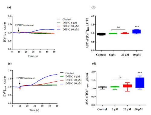
Quantification of the [Ca2+]ER and [Ca2+]cytol levels stimulated by different concentrations of DPHC in EA.hy926 cells. (a,b) The traces (a) and box plots (b) indicating the levels of [Ca2+]ER. (c,d) The traces (c) and box plots (d) indicating the levels of [Ca2+]cytol. For statistical significance, each sample treatment group was compared to the control group. Experiments were performed in triplicates. * p < 0.05, ** p < 0.01, and *** p < 0.001. ns: not significant; AUC: area under the curve; DPHC: diphlorethohydroxycarmalol; [Ca2+]ER: calcium level in the endoplasmic reticulum; [Ca2+]cytol: calcium level in the cytosol; PSS: physiological salt solution. |