
Fig. 10
- ID
- ZDB-FIG-210225-17
- Publication
- Balkrishna et al., 2020 - Calcio-Herbal Medicine Divya-Swasari-Vati Ameliorates SARS-CoV-2 Spike Protein-Induced Pathological Features and Inflammation in Humanized Zebrafish Model by Moderating IL-6 and TNF-α Cytokines
- Other Figures
- All Figure Page
- Back to All Figure Page
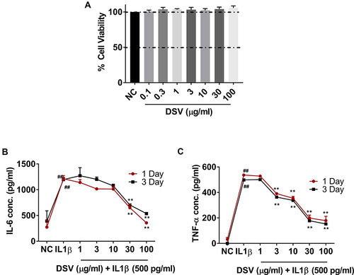
Figure 10 Release of IL-6 and TNF-α from human lung (A549) cells post stimulation with IL-1β in vitro. (A) Cell viability was estimated by Alamar blue assay during a 24-hour period. Results are expressed as mean±SD in triplicate analysis. (B) Cells were pre-treated with different concentration of DSV for either 1 day or 3 days before addition of IL-1β and DSV in the desired concentration. Cell supernatant was used in ELISA for the estimation of IL-6 expression 24 hours post-stimulation. Results are expressed as mean±SD in triplicate analysis. (C) Cell supernatant was used for TNF-α expression as above. Results are expressed as mean±SD ## P<0.001 when compared to the normal control (NC) and ** P<0.01 when compared with the IL-1β stimulated control without any test formulation treatments. |