
Fig. 2
- ID
- ZDB-FIG-210224-79
- Publication
- Widziolek et al., 2020 - Type I interferon-dependent response of zebrafish larvae during tilapia lake virus (TiLV) infection
- Other Figures
- All Figure Page
- Back to All Figure Page
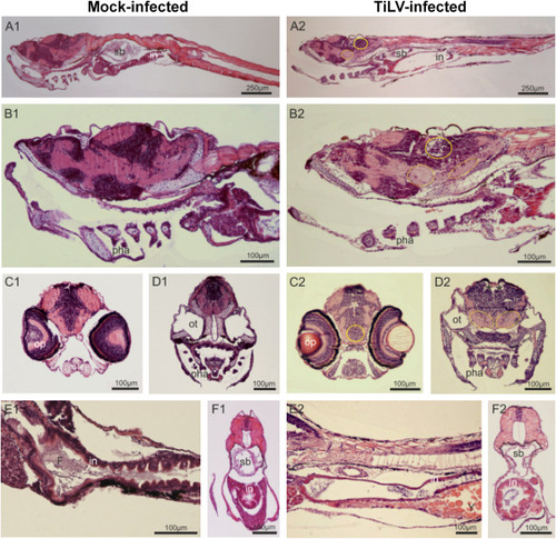
Fig. 2. TiLV-induced histopathological abnormalities in zebrafish larvae. Parasagittal and cross paraffin sections from mock-infected (A1-F1) and TiLV-infected (A2-F2) zebrafish at 4 dpi, stained with H&E. Anterior to the left, dorsal top. N = 6 specimens per each group. In the larvae of TiLV-infected group, all regions of degenerative tissues are encircled in the region of nerve cells bodies and by a dotted oval in the neuropils, where alveolar space is visible. A1-A2: Sagittal section trough larvae till the middle part of the tail (in – intestine; sb – swim bladder). B1–B2: braincase, mouth, gill chamber with pharyngeal arch and gill filaments (pha), anterior part of the digestive tract and liver. C1–C2 and D1-D2: Cross sections of the head on an optic (op) and otic (ot) level. (*) indicates increased blood vessels. E1-E2 and F1–F2: Longitudinal sections through the gastrointestinal region; In mock-infected group intestine epithelium (in) is folded and in the lumen the digested food (F) is present, whereas in TiLV-infected group a delay in gut development is observed with yolk (Y) still present at 6 dpf and minimal folding of intestinal epithelium of some specimens. |