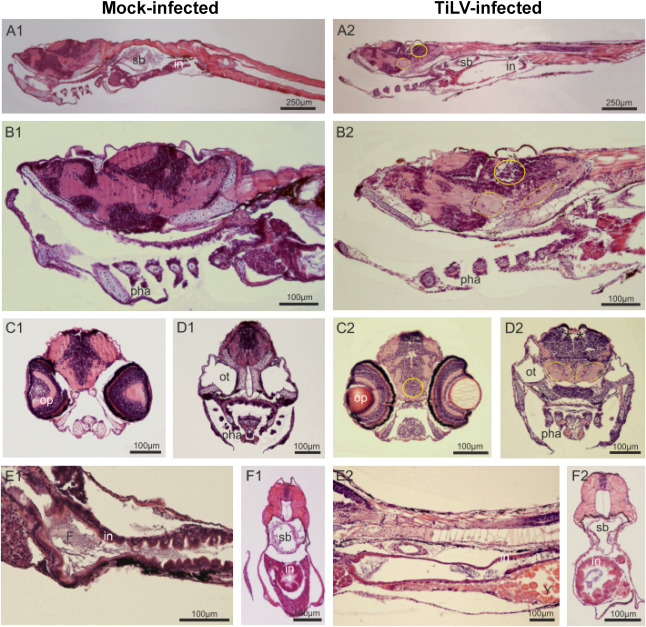
Fig. 2 Fig. 2. TiLV-induced histopathological abnormalities in zebrafish larvae. Parasagittal and cross paraffin sections from mock-infected (A1-F1) and TiLV-infected (A2-F2) zebrafish at 4 dpi, stained with H&E. Anterior to the left, dorsal top. N = 6 specimens per each group. In the larvae of TiLV-infected group, all regions of degenerative tissues are encircled in the region of nerve cells bodies and by a dotted oval in the neuropils, where alveolar space is visible. A1-A2: Sagittal section trough larvae till the middle part of the tail (in – intestine; sb – swim bladder). B1–B2: braincase, mouth, gill chamber with pharyngeal arch and gill filaments (pha), anterior part of the digestive tract and liver. C1–C2 and D1-D2: Cross sections of the head on an optic (op) and otic (ot) level. (*) indicates increased blood vessels. E1-E2 and F1–F2: Longitudinal sections through the gastrointestinal region; In mock-infected group intestine epithelium (in) is folded and in the lumen the digested food (F) is present, whereas in TiLV-infected group a delay in gut development is observed with yolk (Y) still present at 6 dpf and minimal folding of intestinal epithelium of some specimens.
Image
Figure Caption
Acknowledgments
This image is the copyrighted work of the attributed author or publisher, and
ZFIN has permission only to display this image to its users.
Additional permissions should be obtained from the applicable author or publisher of the image.
Full text @ Dev. Comp. Immunol.

