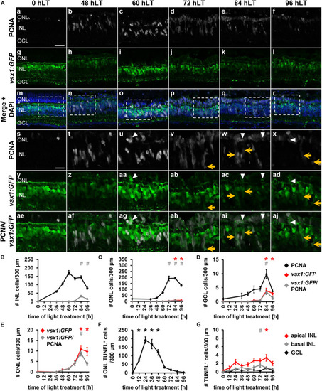
Bipolar cell competence factor vsx1:GFP is expressed in proliferating cells in the light-damaged retina. (Aa–aj) Single z-plane confocal images of retinal sections from light-damaged TgBAC[vsx1:GFP]nns5 zebrafish (0, 48, 60, 72, 84, 96 hLT) immunolabeled for PCNA (Aa–f,Am–x,Aae–aj), GFP (Ag–r,Ay–aj) and counterstained with DAPI (Am–r). (As–aj) Regions outlined in panels (Am–r) at higher magnification. Scale bars, 20 μm (Aa) and 10 μm (As). (B–D) Number of PCNA-positive, vsx1:GFP-positive and PCNA and vsx1:GFP-double positive cells in the INL (B), ONL (C), and GCL (D) over the light treatment timecourse. (E) Number of vsx1:GFP-positive and vsx1:GFP and PCNA−double positive cells in the ONL at a different scale. (F,G) Number of TUNEL-positive cells in the ONL (F) and in the inner retina (G), (apical INL, basal INL, GCL) following constant intense light treatment for 0, 12, 24, 36, 48, 60, 72, 84 and 96 h. Mean ± SE, n ≥ 10. *pTukey < 0.05 and #p < 0.05 indicate comparisons to 0 hLT for the different measures that were assessed. The symbols are color-coded according to the line that they represent in the corresponding graphs (pANOVA, see Table 1). Note, significance was not determined for PCNA in panels (B–D).
|
