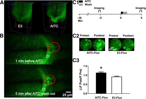
AITC causes an increase in neuronal activity that persists after washout in a hindbrain region of the larval brain. A, Optical recordings of the hindbrain in a GCaMP6 transgenic larva made with a high-speed line scanning confocal fluorescence microscope. 3D reconstructions of a volume of the hindbrain (200 × 140 × 100 μm3) are shown in a larva before and during exposure to AITC. Images were recorded at 5 vols/s at 200 Hz. The images were collected at 1 min before (“E3”) during, and after AITC application. AITC induced strong activation of neurons throughout this brain region. B, Sections from the volume recordings in A. The region of interest (ROI; red circle) was just caudal to the commissura infima Halleri (CI; see Arrenberg et al., 2009). Scale bar, 25 μm. C1, Protocol for examining potential sensitization-related activity in the ROI. Here, activity within the ROI shown in B was imaged for 1 min starting at 6 min before a 30-s exposure to AITC/E3 and at 2.5 min after washout (1 min long) of the AITC. C2, Sample images taken of the ROI before (pretest) and after (posttest) exposure to E3 (images at left) or AITC (images at right). Scale bar, 10 μm. C3, Persistent, postwashout effect of AITC on neuronal activity in the ROI. The normalized fluorescence was significantly greater following exposure to the chemical irritant (AITC-Fluo group, n = 8) than following exposure to E3 (E3-Fluo, n = 8; t(14) = 4.80, p = 0.0003). This figure shows means ± SEM; * indicates a significant (p < 0.05) difference between groups.
|
