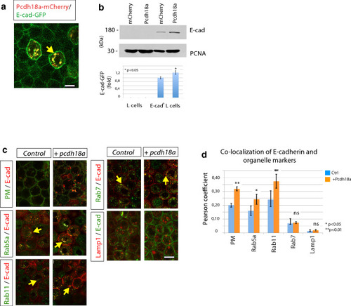
Pcdh18a regulates recycling of the E-cadherin. a Confocal image of zebrafish embryo at 5 hpf. Embryos were microinjected with 0.1 ng of mRNA for the indicated constructs and were imaged in vivo at 5 hpf. Pcdh18a is localized in the cell membrane and in endocytic vesicles, together with E-cadherin (E-cad). b Quantification of the E-cad levels in the Pcdh18a-transfected L cells. Equivalent amounts of lysates from murine L cells or stably E-cadherin-GFP-transfected L cells that had been transfected with Pcdh18a were Western blotted and probed with an anti-GFP antibody; the results showed a 26% increase in the E-cadherin-GFP protein levels after Pcdh18a transfection. The sample blot shows different parts of the same blot and PCNA was used as a loading control. The experiments were performed in independent triplicate (*p value < 0.05; unpaired Student’s t test). c Endocytic routing of E-cad at 50% epiboly. WT embryos and Tg(rab5-GFP), Tg(rab7-GFP), and Tg(rab11-GFP) stable transgenic embryos were microinjected with 0.1 ng of the mRNAs for the indicated constructs. Arrows indicate E-cad localization with Rab proteins and Lamp1-positive vesicles. d Pearson’s co-localization coefficient was calculated from 70 µm thick confocal stacks of five different embryos, each from c. The error bars represent the SEM and significance, as indicated (*p value < 0.05, **p value < 0.01; unpaired Student’s t test). Scale bar: 10 µm
|
