FIGURE

Fig. 3

ID
ZDB-FIG-201113-32
Publication
Moss et al., 2020 - Zebrafish as a model to study autophagy and its role in skeletal development and disease
Other Figures
All Figure Page
Back to All Figure Page
Fig. 3

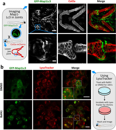
Examples of how GFP-Map1Lc3 transgenic zebrafish line can be used to study autophagy in a skeletal context, a Confocal images of the lower jaw and lower jaw joint of a transgenic CMV:EGFP-map1lc3b zebrafish at 3 days post fertilisation (dpf), immunostained for collagen Type II (red) and anti-GFP for lc3 (green). Scale bar = 50 µm and 10 µm, respectively. b Confocal images of epidermal cells in transgenic CMV:EGFP-map1lc3b zebrafish imaged at 5dpf after treated with 100 µl BafilomycinA1 or DMSO for 16 h followed by 45 min live staining in red LysoTracker dye. Inset boxes show zoom of white checked box. Scale bar = 10 µm

Expression Data

Expression Detail
Antibody Labeling
Phenotype Data

Phenotype Detail
Acknowledgments
This image is the copyrighted work of the attributed author or publisher, and ZFIN has permission only to display this image to its users. Additional permissions should be obtained from the applicable author or publisher of the image. Full text @ Histochem. Cell Biol.