
FIGURE
Figure 1—figure supplement 1.
- ID
- ZDB-FIG-201012-65
- Publication
- Inaba et al., 2020 - Keratins and Plakin family cytolinker proteins control the length of epithelial microridge protrusions
- Other Figures
-
- Figure 1
- Figure 1—figure supplement 1.
- Figure 2
- Figure 3
- Figure 3—figure supplement 1.
- Figure 4
- Figure 4—figure supplement 1.
- Figure 4—figure supplement 2.
- Figure 4—figure supplement 3.
- Figure 4—figure supplement 4.
- Figure 4—figure supplement 5.
- Figure 4—figure supplement 6.
- Figure 4—figure supplement 7.
- Figure 5
- Figure 5—figure supplement 1.
- Figure 6.
- Figure 7.
- Figure 8.
- All Figure Page
- Back to All Figure Page
Figure 1—figure supplement 1.
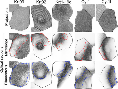
GFP-tagged keratin BAC reporters expressed in periderm cells at 48hpf. The first row shows maximum intensity projections, the second and third rows show optical sections highlighting the microridge-like pattern at the apical surface of cells (red outlines) or the filamentous pattern within the same cells (blue outlines). Dashed lines indicate the outlines of individual periderm cells. Images were inverted so that high-intensity fluorescence appears black and low-intensity fluorescence is white. |
Expression Data
Expression Detail
Antibody Labeling
Phenotype Data
Phenotype Detail
Acknowledgments
This image is the copyrighted work of the attributed author or publisher, and
ZFIN has permission only to display this image to its users.
Additional permissions should be obtained from the applicable author or publisher of the image.
Full text @ Elife