|
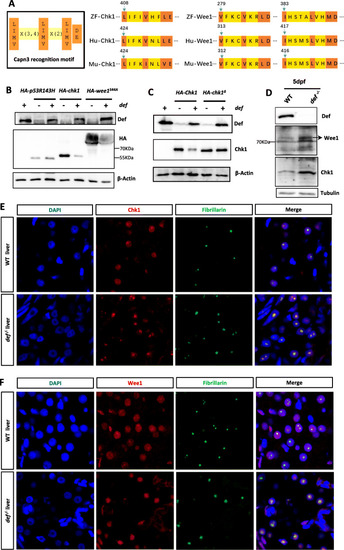
Chk1 and Wee1 are substrates of the Def-Capn3b complex. a Diagram showing the Capn3 recognition motif (left panel) and the locations of this motif in zebrafish, human and mouse Chk1 (middle panel) and Wee1 (right panel) proteins. b, c Western blot of Def, HA-p53R143H, HA-Chk1 and HA-Wee1S44A in WT embryos injected with HA-p53R143H, HA-Chk1 or HA-Wee1S44A mRNA combined with or without def mRNA (b). Western blot of HA-Chk1 and HA-Chk1Δ in WT embryos injected with HA-Chek1or HA-Chek1Δ mRNA combined with or without def mRNA (c). Total proteins were extracted at 8-h post-injection. β-Actin: loading control. d Western blot of the endogenous Chk1 and Wee1 proteins in WT and def−/− mutant embryos at 5dpf. Tubulin: loading control. e, f Co-immunostaining of Chk1 (e) or Wee1 (F) with Fibrillarin in the liver of WT with def−/− at 5dpf. DAPI: stain nuclei
|