FIGURE

FIGURE 1

ID
ZDB-FIG-200614-18
Publication
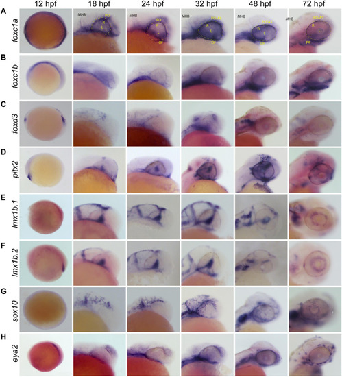
Van Der Meulen et al., 2020 - Spatiotemporal Characterization of Anterior Segment Mesenchyme Heterogeneity During Zebrafish Ocular Anterior Segment Development
Other Figures
All Figure Page
Back to All Figure Page
FIGURE 1

Whole Mount In Situ Hybridization (WISH) of known POM and neural crest-related marker genes. Whole-mount in situ hybridization for POM marker gene mRNA expression patterns were observed during early to late stage eye development in the lateral view. POM genes examined were (A)foxc1a, (B)foxc1b, (C)foxd3, (D)pitx2, (E)lmx1b.1, (F)lmx1b.2, (G)sox10, and (H)eya2. Foxc1a, foxc1b, pitx2, eya2, and sox10 in particular show strong expression surrounding the optic cup and on the surface of the anterior segment from 24 to 72 hpf. Lmx1b.1 and lmx1b.2 expression is detected within the anterior segment, surrounding the lens by 48–72 hpf. MHB, midbrain-hindbrain boundary; R, retina; L, lens; OV, optic vesicle; PO, periocular space (outlined with dashed line); OF, optic fissure; AS, anterior segment.

Expression Data

Expression Detail
Antibody Labeling
Phenotype Data

Phenotype Detail
Acknowledgments
This image is the copyrighted work of the attributed author or publisher, and ZFIN has permission only to display this image to its users. Additional permissions should be obtained from the applicable author or publisher of the image. Full text @ Front Cell Dev Biol