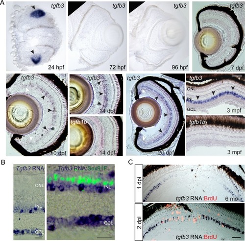
(A) tgfb3 in situ hybridization identifies tgfb3 expression in lens at 24 hpf (hours post fertilization) and in MG beginning ~10 dpf (days post fertilization). This latter expression continues to increase throughout the first 3 months of development. Note tgfb1b expression remains undetectable by in situ hybridization at all the time points examined (14 dpf and 3 mpf). Size marker is 20 microns. In the 10 and 20 dpf panels, black arrowheads point to tgfb3expressing MG in the central retina, while white arrowheads point to reduced tgfb3 expression in the retinal periphery. (B) In situ hybridization (blue/purple product) and Sox9 immunofluorescence (green fluorescence) identifies tgfb3 expression in GCL and INL, but not in MG of the mouse retina. Size marker is 25 microns. (C) Retinal injury suppresses tgfb3expression at the injury site in 3 month old fish. In situ hybridization detects tgfb3 RNA (blue/purple product) and BrdU immunofluorescence (red/orange fluorescence) identifies proliferating cells. Size marker is 40 microns.