
Fig. 3 S1
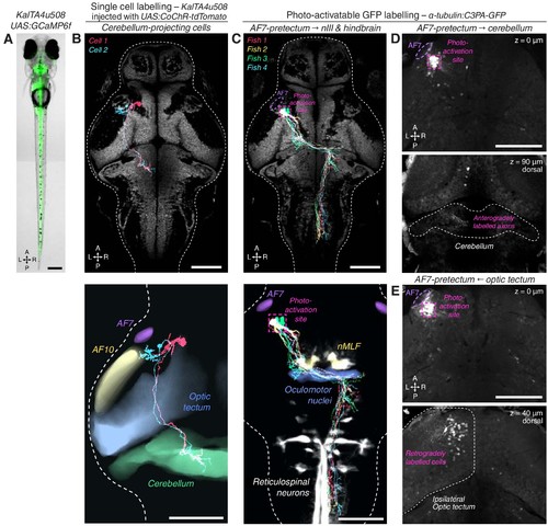
KalTA4u508 neurons innervating cerebellum, and PA-GFP projection mapping from AF7-pretectum.(A) Expression pattern of the KalTA4u508 transgene, illustrated by a KalTA4u508;UAS:GCaMP6ftransgenic larva (6 dpf). (B) Tracings of KalTA4u508 neurons projecting to ipsilateral medial corpus cerebellum (N = 2 cells from two fish). The bottom image shows tracings overlaid with selected anatomical regions from the ZBB brain atlas. (C) Tracings of PA-GFP-labelled AF7-pretectal cells projecting to oculomotor nuclei and contralateral hindbrain in 7 dpf α-tubulin:C3PA-GFP larvae (N = 4 fish) registered to the elavl3:H2B-GCaMP6s reference brain (grey). The photo-activation site is indicated in magenta. (D) PA-GFP-labelled AF7-pretectal cells in a 7 dpf α-tubulin:C3PA-GFP larva. The photo-activation site is indicated in magenta. Anterogradely labelled axonal terminals are visible in the ipsilateral medial cerebellum (bottom image, z-plane location is relative to top z-plane). (E) A second example of photoactivation that retrogradely labelled cell bodies in the ipsilateral anterior-ventral optic tectum. Scale bars, 100 µm except (A), 200 µm. A, anterior; L, left; P, posterior; R, right. |