FIGURE

Fig. 7

ID
ZDB-FIG-200501-7
Publication
Vlasschaert et al., 2015 - Evolution of the highly networked deubiquitinating enzymes USP4, USP15, and USP11
Other Figures
All Figure Page
Back to All Figure Page
Fig. 7

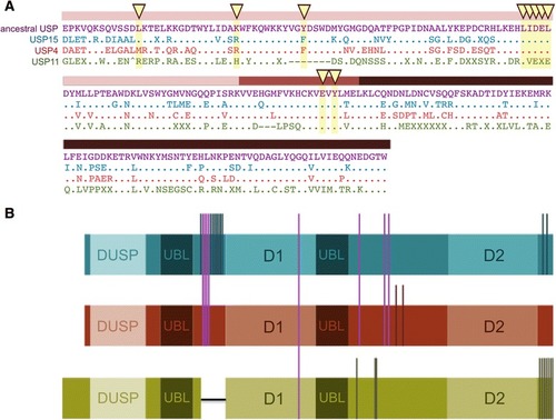
Summary of signature features of USP4, USP15 and USP1. a Alignment of signature sequences for USP4 (red), USP15 (blue) and USP11 (green) with the single-copy sequence (purple). The DUSP-UBL compound domain is shown and coloured as in Fig. 1. b Schematic illustration of signature phosphorylation sites and loss of alternatively spliced exon in USP11. Vertical bars traverse sequences with shared phosphorylation sites

Expression Data

Expression Detail
Antibody Labeling
Phenotype Data

Phenotype Detail
Acknowledgments
This image is the copyrighted work of the attributed author or publisher, and ZFIN has permission only to display this image to its users. Additional permissions should be obtained from the applicable author or publisher of the image. Full text @ BMC Evol. Biol.