|
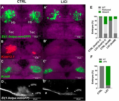
Premature activation of Wnt signaling causes a reduction of dHbl neurons. (A-D′) Projections of confocal z-stacks. (A-C′) Dorsal views, anterior is towards top focused onto the diencephalon of (A-D′) embryos at 3 dpf. Nuclei are DAPI labeled (purple). (A,A′) LiCl treatment causes delayed habenular neuron differentiation. (B-C′,E) Markers for (B,B′) dHbl and (C,C′) dHbm neurons are reduced as represented in E. (D,D′) Lateral views of IPN innervation by habenular efferent axons, anterior is leftwards. Treated embryos show vIPN innervation, as represented in F. d, dorsal; Hb, habenula, IPN, interpeduncular nucleus; Tec, optic tectum; v, ventral.
|