FIGURE

Figure 1

ID
ZDB-FIG-200421-72
Publication
Giribaldi et al., 2020 - Synthesis, Pharmacological and Structural Characterization of Novel Conopressins from Conus miliaris
Other Figures
All Figure Page
Back to All Figure Page
Figure 1

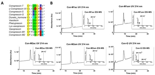
RP-HPLC/ESI-MS analyses of the synthesized conopressins and alignment of conopressin-related sequences. (A) Alignment of conopressin-related sequences. The asterisks * indicate an amidated C-terminal. Conopressin-M1 and M2 with γ-conopressin-vil are the only sequences that display a negatively charged amino acid at position 8. Interestingly, conopressin-M1 also displays an unusual proline residue at position 3. The highly conserved glycine residue at position 9 is replaced by a serine residue in conopressin-M1 and M2. (B) RP-HPLC/ESI-MS analyses of the synthesized conopressins. Acetonitrile (ACN) gradient from 0% to 30% over 30 min. For Con-M1 the two peaks display the same mass, possibly caused by the two proline residues inducing cis-trans isomerization causing dynamic conformational exchange leading to the splitting of the UV chromatogram peak [16,17,18]. The asterisk (*) on ESI-MS insets indicate an ion resulting from in source fragmentation of the proline residue [19].

Expression Data

Expression Detail
Antibody Labeling
Phenotype Data

Phenotype Detail
Acknowledgments
This image is the copyrighted work of the attributed author or publisher, and ZFIN has permission only to display this image to its users. Additional permissions should be obtained from the applicable author or publisher of the image. Full text @ Mar. Drugs