|
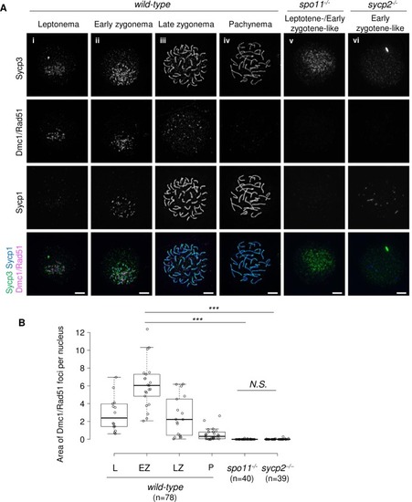
Formation of Dmc1/Rad51 foci is impaired in <italic>sycp2</italic><sup><italic>-/-</italic></sup> spermatocytes.A: Immunostaining of Dmc1/Rad51, Sycp1 and Sycp3 on wild-type (i to iv), spo11-/- (v) and sycp2-/- (vi) spermatocyte chromosomal spreads. The wild-type nuclei are at leptonema (i), early zygonema (ii), late zygonema (iii) and pachynema (iv) according to the Sycp1 and Sycp3 staining patterns. A spo11-/- nucleus at an early zygotene-like stage, according to Sycp3 staining patterns, is shown (v). An early zygotene-like sycp2-/- nucleus stained with short Sycp1 fragments is shown (vi). Scale bars, 5 μm. B: Quantification of the Dmc1/Rad51-stained area. The sum of the area stained for the Dmc1/Rad51 foci in each nucleus was measured in wild-type nuclei at leptonema (L, n = 14), early zygonema (EZ, n = 21), late zygonema (LZ, n = 17), and pachynema (P, n = 26), in leptotene- or early zygotene-like spo11-/- (n = 40), and in early zygotene-like sycp2-/- (n = 39) spermatocytes. Center lines show the medians; box limits indicate the 25th and 75th percentiles as determined by R software; whiskers extend 1.5 times the interquartile range from the 25th and 75th percentiles; data points are plotted as open circles. Chromosomal spreads of two individual fish were used for each genotype. *** indicates p<0.0001 (Student’s t-test). N.S. indicates not significant.
|