
FIGURE
Figure 5
- ID
- ZDB-FIG-200325-46
- Publication
- Dang et al., 2020 - Evolutionary and Molecular Characterization of liver-enriched gene 1
- Other Figures
- All Figure Page
- Back to All Figure Page
Figure 5
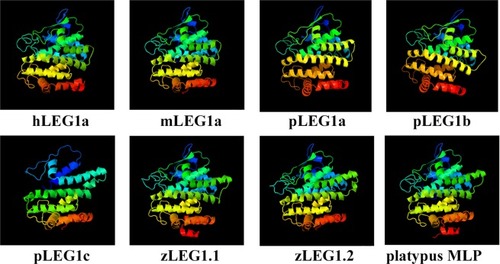
Structural comparison of LEG1 proteins from different species. The platypus MLP protein structure was retrieved from the PDB (4V00), while the others were predicted using Phyre 2. The colors are in rainbow order with red and blue colors indicate the N- and C- termini of LEG1, respectively. All LEG1 proteins exhibit the similar structural prediction result expect for pLEG1c, which is slightly different from others due to the lack of the signal peptide. |
Expression Data
Expression Detail
Antibody Labeling
Phenotype Data
Phenotype Detail
Acknowledgments
This image is the copyrighted work of the attributed author or publisher, and
ZFIN has permission only to display this image to its users.
Additional permissions should be obtained from the applicable author or publisher of the image.
Full text @ Sci. Rep.