
Fig. 5
- ID
- ZDB-FIG-200325-4
- Publication
- Wang et al., 2019 - The Application of Methylprednisolone Nanoscale Zirconium-Porphyrin Metal-Organic Framework (MPS-NPMOF) in the Treatment of Photoreceptor Degeneration
- Other Figures
- All Figure Page
- Back to All Figure Page
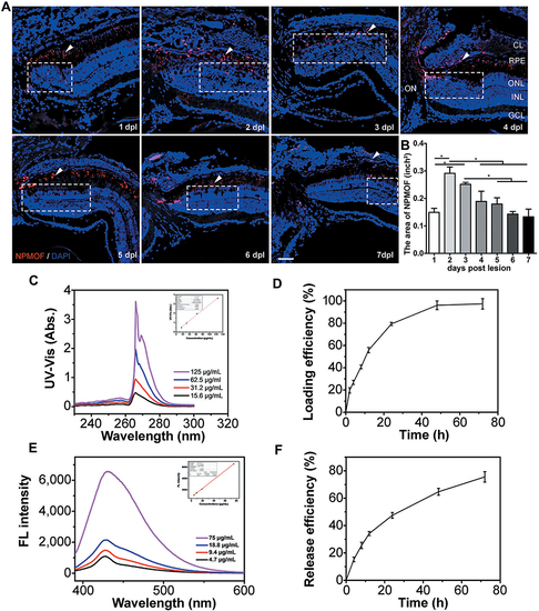
Distribution of NPMOF in the retina from 1 to 7 days post lesion and the loading efficiency and release efficiency for MPS in NPMOF. (A) The distribution of NPMOF (arrowheads) around the lesioned area (dotted rectangles) in the retina from 1 to 7 dpl. (B) The statistical analysis of NPMOF in the retina (ANOVA, *p<0.05). (C and D) The UV-Vis absorbance (insert, standard curve) of MPS in DMF and its loading performance. (E and F) The fluorescence (insert, standard curve) of MPS in 5% hyaluronic acid and its release performance. Scale bar in (A): 50 μm. Abbreviations: CL, choroid layer; RPE, retinal pigment epithelium; ONL, outer nuclear layer; INL, inner nuclear layer; GCL, ganglion cell layer; ON, optic nerve. |