
Figure 5
- ID
- ZDB-FIG-200228-5
- Publication
- Einfalt et al., 2020 - Bioinspired Molecular Factories with Architecture and In Vivo Functionalities as Cell Mimics
- Other Figures
- All Figure Page
- Back to All Figure Page
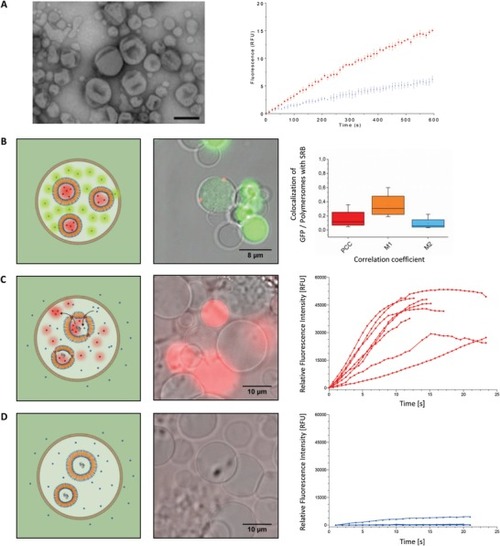
Engineering a MF. A) Left: TEM micrograph of AOs based on PMOXA6‐PDMS44‐PMOXA6 polymersomes loaded with horseradish peroxidase (HRP) and equipped with OmpF. Scale bar: 500 nm. Right: Amplex UltraRed conversion kinetics of AOs equipped with OmpF (red) and AOs without OmpF (blue). Standard deviations are based on 3 individual measurements. B) Spatial colocalization in E‐GPMVs. Left: Schematic representation of E‐GPMVs containing SRB‐loaded polymersomes and cytosolic GFP protein. Middle: CLSM micrograph of E‐GPMVs containing SRB‐loaded polymersomes (Red signal) and cytosolic GFP protein (Green signal). Right: Colocalization analysis of SRB‐loaded polymersomes and GFP protein within E‐GPMVs. PCC = 0.16 ± 0.14, M1 = 0.35 ± 0.1, M2 = 0.09 ± 0.09. |