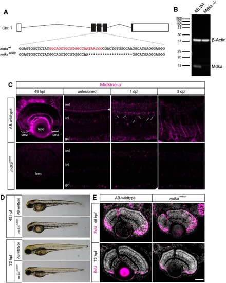
Midkine-a governs cell cycle kinetics during retinal development. A, Schematic representation of the midkine-a locus on chromosome 7. The gene consists of five exons. Red represents the gRNA target sequence located at exon 3. B, Using larvae at 48 h post fertilization (hpf), Western blot analysis for Midkine-a confirms lack of protein in the mdkami5001 mutant. C, Immunocytochemistry for Midkine-a in embryonic (48 hpf), adult, and regenerating retinas. In unlesioned retina, Midkine-a immunoreactivity is detected in horizontal cells (arrowhead) (Gramage et al., 2015). Photoreceptor cell death induces Midkine-a in reprogrammed Müller glia (arrows) at 1 dpl. Midkine-a distributes radial processes of Müller glia at 3 dpl. D, Lateral views of larvae at 48 and 72 hpf: AB-WT and mdkami5001 mutant. E, Proliferation assay with EdU labeling at 48 hpf. Compared with WT, there is an increased number of EdU-labeled cells in the retinas of mdkami5001 mutants. Retinal lamination and cellular differentiation are delayed, but not blocked in the mdkami5001 at 72 hpf. cmz, Ciliary marginal zone; onl, outer nuclear layer; inl, inner nuclear layer; gcl, ganglion cell layer. Scale bar, 40 μm. (see extended data, Table 1-1, and Table 1-2).
|
