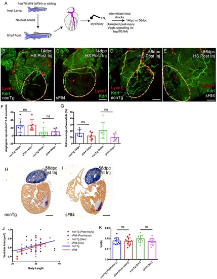
Induction of sFlt4 after cryoinjury has no significant effect on coronary vessels or heart morphology.
(A) Schematic representation of the experimental inhibition of post-injury signaling through lymphatic endothelial receptor Flt4. Whole-mount confocal (B–E) and brightfield AFOG-histology imaging (H and I) of post-injury heat shocked non-transgenic (B, D, H), hsp70l:sflt4 (C, E, I) adult transgenic zebrafish hearts that express arterially and endocardially enriched endothelial marker kdrl:GFP (green: B-E), lymphatic endothelial marker lyve1:RFP (red: B-E). (B and C) Hearts from both hsp70l:sflt4and non-transgenic post-injury heat shocked zebrafish show angiogenic sprouts and formed vessels within the woundsite (demarked in orange) at 14dpc. (D and E) Hearts from both hsp70l:sflt4 and non-transgenic post-injury heat shocked zebrafish show increased coronary vessel coverage by 56dpc. (F) Quantification of normalized coronary vessel angiogenic sprouts at 14 and 56dpc in the wound site of hearts from both hsp70l:sflt4 and non-transgenic post-injury heat shocked zebrafish. (G) Quantification of coronary vessel coverage of the wound site at 14 and 56dpc in hearts from both hsp70l:sflt4 and non-transgenic post-injury (HS 242dpf, cryoinjury 244dpf) heat shocked zebrafish. (H and I) Example images of largest AFOG stained tissue section used for ventricle quantification. (J) Linear regression analysis showing increasing max ventricle area to body length of AFOG-stained nonTg and hsp70l:sflt4 zebrafish heart sections with either post-injury heat shock (14 and 56dpc, HS from 242dpf, cryoinjured at 244dpf, see A) or developmental heat shock (Dev; 21dpc or 60dpc, HS from 35 to 205dpf or 71dpf to 229dpf, cryoinjury at 206 or 230dpf, see Figure 6D). (K) Quantification of max ventricle area (VA) to body length (BL) of nonTg and hsp70l:sflt4 zebrafish (post-injury HS: 14 and 56dpc and developmental (Dev) HS: 21 and 60dpc) showing no significant effect on post injury ventricle size with sFlt4 induction.
Source data for Supplement 3(<bold>F</bold>) and (<bold>G</bold>).
Expression Data
Expression Detail
Antibody Labeling
Phenotype Data
Phenotype Detail
Acknowledgments
This image is the copyrighted work of the attributed author or publisher, and
ZFIN has permission only to display this image to its users.
Additional permissions should be obtained from the applicable author or publisher of the image.
Full text @ Elife
Your Input Welcome
Thank you for submitting comments. Your input has been emailed to ZFIN curators who may contact you if
additional information is required.
Oops. Something went wrong. Please try again later.