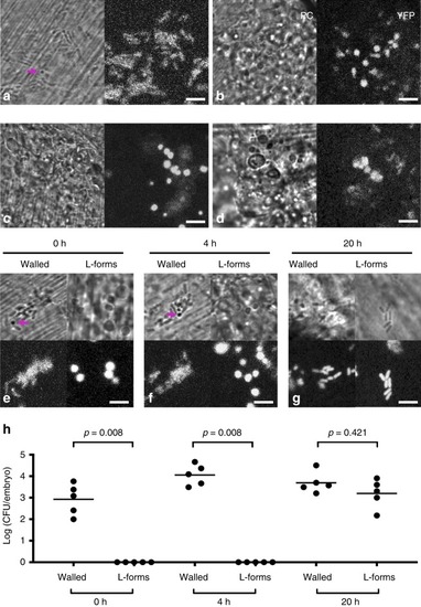
L-form switching in the zebrafish embryo. a, b Walled E. coli ST144-YFP was injected into the tail fin of zebrafish larvae in the presence (b) or absence (a) of 0.4 mg/ml phosfomycin and visualised by phase contrast (PC) or fluorescence microscopy (YFP). The magenta arrow in (a) points to a bacterium that appears to have adopted a round shape in the absence of antibiotic. c, d ST144-YFP E. coli L-forms induced in vitro with 0.4 mg/ml phosfomycin were injected into the tail fins of zebrafish larvae in the presence of 0.4 mg/ml phosfomycin and visualised by microscopy immediately post-injection (c), or following an overnight incubation (d). e–g ST144-YFP E. coli walled or in vitro induced L-form bacteria were injected in the absence of the antibiotic and visualised 0 h (e), 4 h (f) or 20 h (g) post infection. h The bacteria were enumerated by homogenising fish embyos and plating out on NA 0, 4 or 20 h post infection. Each circle represents recovered bacteria from an individual larva. Representative data from two independent experiments (one with two and one with three larvae). Mean (horizontal bars) is shown. The p values were determined by non-parametric Mann–Whitney test. Significance was defined as p < 0.05. Scale bars = 5 µm. Source data for Fig. 5h are provided as Source Data file
|
