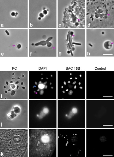
L-form-like structures observed in the urine of various patients. a–h Examples of fresh urine samples assessed by phase contrast microscopy. Bundels typical of dividing L-forms (a, b). Magenta arrows in (c) and (d) point to putative L-form bacteria associated with sluffed eukaryotic cells; in (e) and (f) they point at crescent shaped bulges, reminiscent of the outer membrane in L-forms of Gram-negative bacteria; in (g) a possible intermediate stage between walled and L-form is marked; and in (h), internal membrane vesicles are evident. i–k Examples of fixed samples assessed by fluorescence in situ hybridisation (FISH). Patient samples were fixed with paraformaldehyde and stained with DAPI and a fluorescently labelled DNA probe against bacterial 16S rRNA (BAC 16S). To show the specificity of DAPI and the probe staining an image was acquired in the red channel, which did not produce significant fluorescence (Control). i Two L-form-like structures were observed among several likely walled bacteria. One of the objects stained both with the bacterial probe and DAPI (magenta arrow) while the other one, only with DAPI (blue arrow). j Example of L-form-like structures of varied size that stained with the bacterial probe. k Example showing a eukaryotic cell in urine in the same field of view as numerous bacteria. The bacterial DNA and the nucleous of the eukaryotic cell (n) both stained with DAPI but the BAC 16S probe only associated with the bacterial cells, demonstrating the specificity of the probe. Scale bars = 5 µm, apart form panel (k), where the scale bar = 15 µm
|
