
Fig. 6
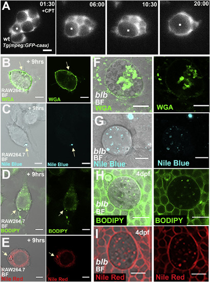
In Macrophages and Microglia, the Gastrosome Contains Lipid and Membranes (A) SPIM time-lapse of a representative 3 dpf wild-type Tg(mpeg:GFP-caax) microglia in an embryo treated with Camptothecin to increase apoptosis. Scale bar 5 μm. The while dot marks the growing compartment. See also Video S6. (B–E) RAW264.7 macrophages fixed 9 h after apoptosis induction of unlabeled HeLa cells. Stained with WGA (B), Nile Blue (C), Bodipy (D), and Nile Red (E). The white arrow points to the gastrosome. Scale bar, 5 μm. (F–I) 4 dpf blb microglia stained with WGA (F), Nile Blue (G), Bodipy (H) and Nile Red (I). Scale bar, 10 μm. |
| Gene: | |
|---|---|
| Fish: | |
| Condition: | |
| Anatomical Terms: | |
| Stage: | Protruding-mouth |
| Fish: | |
|---|---|
| Condition: | |
| Observed In: | |
| Stage: | Protruding-mouth |
Reprinted from Developmental Cell, 49(1), Villani, A., Benjaminsen, J., Moritz, C., Henke, K., Hartmann, J., Norlin, N., Richter, K., Schieber, N.L., Franke, T., Schwab, Y., Peri, F., Clearance by Microglia Depends on Packaging of Phagosomes into a Unique Cellular Compartment, 77-88.e7, Copyright (2019) with permission from Elsevier. Full text @ Dev. Cell