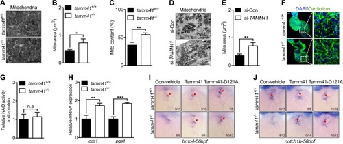
Tamm41 deficiency leads to cardiomyocytes mitochondrial defects. a–c TEM images show enlarged mitochondria (lower panel) in the heart tissues of tamm41−/− embryos compared with mitochondria (upper panel) in tamm41+/+ cardiomyocytes. b and c are quantitative data for mitochondrial areas and mitochondrial contents in tamm41+/+ and tamm41−/− hearts based on TEM images respectively. d, e TEM analysis of human cardiomyocyte cell line (AC16) reveals enlarged mitochondria formation with TAMM41 deficiency. Mitochondrial area quantitation is shown in (e). f Immunofluorescence analysis of cardiolipin revealed no obvious differences in fluorescence signals between tamm41+/+ and tamm41−/− hearts. Right are the enlarged ones. Scale bar: 20 μm. g Relative CL level assessed by NAO in mitochondria isolated from 52hpf tamm41+/+ and tamm41−/− embryos. h Elevated cardiolipin synthesis-related genes (cds1 and pgs1) in tamm41−/− embryos. i, j WISH assessment of valve-related markers with tamm41, tamm41-D121A mRNA overexpression. Tamm41-D121A, which has been suggested defective in cardiolipin synthesis, harbors the same effects in restoring the heart valves of tamm41−/− embryos (third panel in i and j, red arrows) as with tamm41 overexpression (second panel in i and j, red arrows). Black horizontal lines indicate mean ± SD. Means ± SD are shown for three independent experiments. n.s, not significant, *P < 0.05; **P < 0.01; ***P < 0.001 (Student’s t-test)
|
