
FIGURE
Fig. 1
- ID
- ZDB-FIG-190827-28
- Publication
- Monma et al., 2019 - Aging-associated microstructural deterioration of vertebra in zebrafish
- Other Figures
- All Figure Page
- Back to All Figure Page
Fig. 1
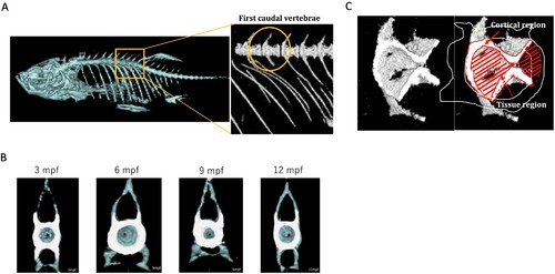
Morphological images with micro-CT visualisation. A. The position of FCV. FCV exhibits elongated unfused haemal arches with shortened ribs, and absence of a haemal spine (Bird and Mabee, 2003). We selected this vertebra in the following analyses. B. Cross-sectional images of FCV in 3–12 mpf zebrafish. Surrounding bone over the threshold of 300 mg/cm3 in BMD is defined as the cortical bone (white area), and the region surrounded by cortical bone is defined as ‘tissue’ (blue area), containing trabecular bone. C. Representative 3-D images of FCV. |
Expression Data
Expression Detail
Antibody Labeling
Phenotype Data
Phenotype Detail
Acknowledgments
This image is the copyrighted work of the attributed author or publisher, and
ZFIN has permission only to display this image to its users.
Additional permissions should be obtained from the applicable author or publisher of the image.
Full text @ Bone Rep